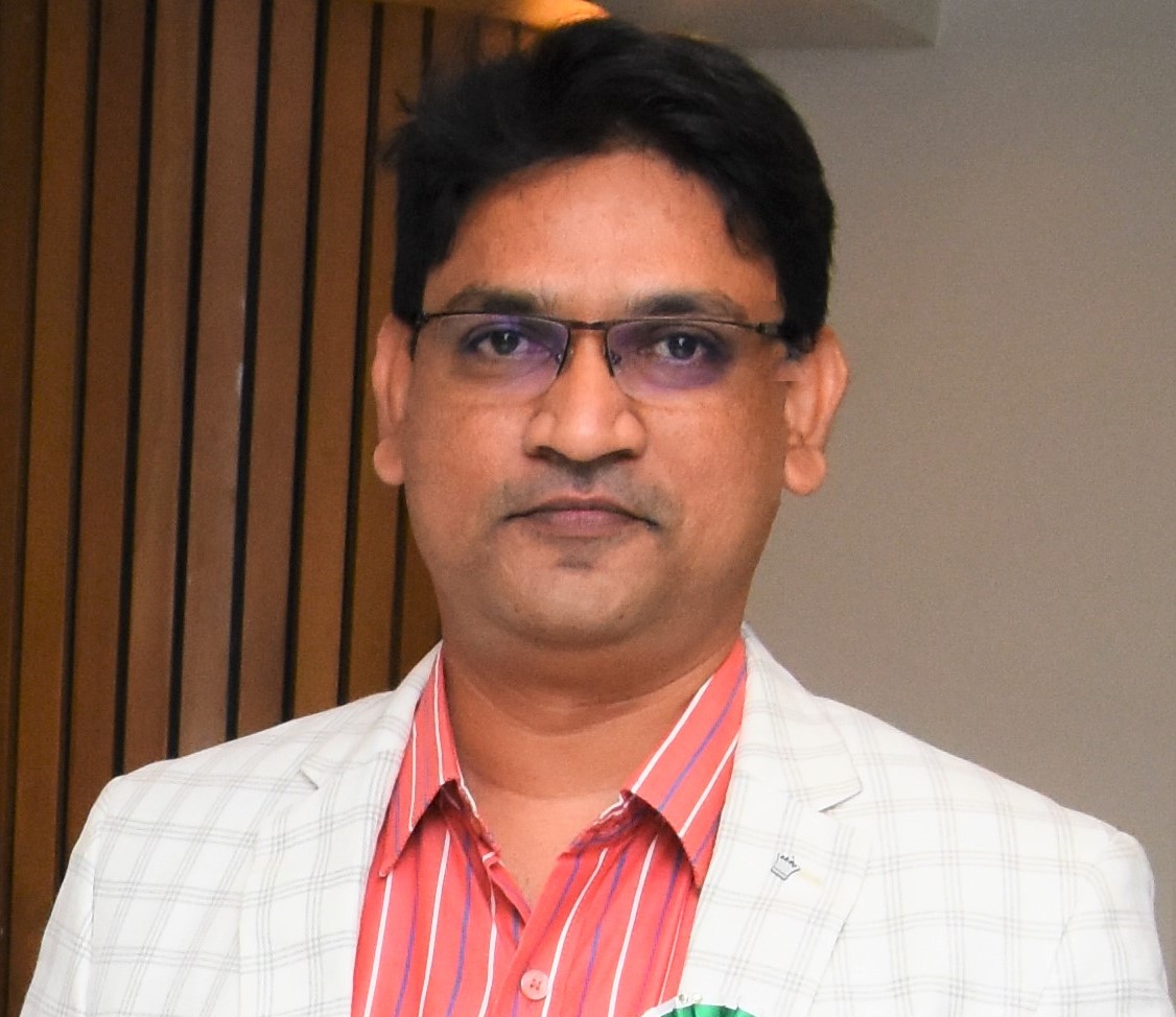
Dr.V VIJAYA KISHORE
@gpcet.ac.in
PROFESSOR, ELECTRONICS AND COMMUNICATION ENGINEERING
G PULLAIAH COLLEGE OF ENGINEERING AND TECHNOLOGY, KURNOOL, AP,INDIA
Dr. V. Vijaya Kishore is a Professor in department of Electronics and Communication Engineering at G Pullaiah College of Engineering and Technology, Kurnool, AP. He has a teaching experience of 18+ years. He holds Ph.D. degree in Electronics and Communication Engineering department from S.V.University, Tirupati in the field of Bio-medical Image Processing. He has published more than 35 research papers in journals of both international and national repute. He published two articles on Effective Engineering Teaching. He has successfully completed the IITBombayX Foundation Program in ICT for Education, FDP101X and FDP201X. He has published two books in the area of Biomedical Image Processing. His research areas are Biomedical Image Processing, Computer Aided Diagnosis tools development. His main focus is on developing tools that help for segmentation and extraction of region of interest in medical images and for decision making on the diagnosis. He received National award of Excellence in teaching from Global Management Council. He is acting as reviewer for four SCI journals and one Scopus journal related to Medical and Image Processing. He is also a member of Editorial Review Board of International Journal of Biomedical and Clinical Engineering journal. He has been invited as Session chair and Program Committee Member for various national and international conferences.
RESEARCH INTERESTS
MEDICAL IMAGE PROCESSING, BIOMEDICAL SIGNAL PROCESSING, COMPUTER AIDED DETECTION TOOL DESIGN AND DEVELOPMENT, IMAGE PROCESSING.
Scopus Publications
Scopus Publications
Pasupuleti Naga Sudhakar and V. Vijaya Kishore
Elsevier BV
Vijaya Kishore Veparala and Vattikunta Kalpana
Salud, Ciencia y Tecnologia
En la era actual de las tecnologías de la información, la información es el factor más importante para determinar cómo progresarán los distintos paradigmas. Esta información debe extraerse de un enorme tesoro informático. El aumento de la cantidad de datos analizados e interpretados es consecuencia directa de la proliferación de plataformas de procesamiento más potentes, el incremento del espacio de almacenamiento disponible y la transición hacia el uso de plataformas electrónicas. En este trabajo se describe un estudio exhaustivo de Big Data, sus características y el papel que desempeña el algoritmo de clustering Subspace. La contribución más importante que hace este trabajo es que lee muchas investigaciones anteriores y luego hace una presentación exhaustiva sobre las diferentes formas en que otros autores han clasificado los métodos de clustering subespacial. Además, se han proporcionado, con una breve explicación, algoritmos significativos que pueden servir de referencia para cualquier desarrollo futuro.
V Vijaya Kishore, V Kalpana, and M Jayalakshmi
IEEE
Data mining has become increasingly important in recent years for the capacity of the medical industry to anticipate illness outbreaks. The process of selecting, analyzing, and modelling massive amounts of dossier information with the goal of locating previously unknown connections or alliances that are significant to information researchers is known as “data mining.” Data mining is a technique. Diabetes is a condition that is induced by having an abnormally high amount of glucose fixation in the blood. Several different computational understanding systems that use various classifications to forecast and identify diabetes were explained. The choice of dependable classifiers unquestionably contributes to an increase in the precision and proficiency of the system. In this article, a technique is proposed that identifies data by utilizing an SVM classifier that has been altered. Within the scope of this investigation, we developed a computational model with the goal of improving diabetes forecasting.
V. Kalpana, V. Vijaya Kishore, and B. Hari Krishna
Institution of Engineering and Technology
T. Tirupal, Y. Pandurangaiah, Ajay Roy, V. Vijaya Kishore, and Anand Nayyar
Springer Science and Business Media LLC
V. Vijaya Kishore, Shilpa Sreya Batthala, Jithendra Varma Chamarthi, Chalambu Achyutasai, and B Subrahmanyam
Faculty of Engineering, University of Kragujevac
Sruthy R, S. Kavitha, N. Darwin, Anita Titus, V. Vijaya Kishore, and Dharshini. B. S
IEEE
While old techniques are time-consuming and inefficient, recent years have seen a rise in the importance of student attendance as a reflection of academic accomplishments and the effectiveness provided to any university. Recently, however, a variety of automated identifying technologies, such as Radio Frequency Identification (RFID), have gained popularity (RFID). Many studies and applications being developed to make the most of this technology, which raises certain ethical questions. RFID, or radio-frequency identification, is a wireless technology used for the purpose of identifying and monitoring an object by the transfer of data from an electronic tag, termed an RFID tag or label, via radio waves to an RFID reader. In this project, an RFID-based system has been developed to provide an attendance monitoring system. In addition to streamlining the process as a whole, automated attendance management software will also produce a well-structured and analyzed report of the pattern of student attendance and time management, which may aid in the allocation and use of human resources. In this study, RFID was used to the task of tracking student attendance, allowing teachers and administrators to more efficiently record in-person classroom statistics that may be used to determine how students should be graded and inform other administrative choices.
K. Maheswari, M. L. Ravi Chandra, D. Srinivasulu Reddy, and V. Vijaya Kishore
FOREX Publication
This work presents a novel technique to develop the three-valued logic (TVL) circuit schematics for very large-scale integration (VLSI) applications. The TVL is better alternative technology over the two-valued logic because it provides decreased interconnect connections, fast computation speed and decreases the chip complexity. The TVL based complicated designs such as half-adder and multiplier circuits are designed utilizing the Pseudo N-type carbon nanotube field effect transistors (CNTFETs). The proposed TVL half adder multiplier schematics are developed in HSPICE tool. Additionally, the delay and circuit area for the half- adder and multiplier circuits are investigated and compared to the complementary circuits. The memory usage and CPU time for the proposed circuits are also analyzed. It is observed that the proposed circuit designs show the improved performance up to 43.03% on an average over the complementary designs.
V. Kalpana, V. Vijaya Kishore, and R. V. S. Satyanarayana
Springer Nature Singapore
V. Vijaya Kishore and R.V.S. Satyanarayana
IGI Global
A vital necessity for clinical determination and treatment is an opportunity to prepare a procedure that is universally adaptable. Computer aided diagnosis (CAD) of various medical conditions has seen a tremendous growth in recent years. The frameworks combined with expanding capacity, the coliseum of CAD is touching new spaces. The goal of proposed work is to build an easy to understand multifunctional GUI Device for CAD that performs intelligent preparing of lung CT images. Functions implemented are to achieve region of interest (ROI) segmentation for nodule detection. The nodule extraction from ROI is implemented by morphological operations, reducing the complexity and making the system suitable for real-time applications. In addition, an interactive 3D viewer and performance measure tool that quantifies and measures the nodules is integrated. The results are validated through clinical expert. This serves as a foundation to determine, the decision of treatment and the prospect of recovery.
The 2021 International Conference on Power Electronics and Power Transmission (ICPEPT 2021) was held on October 15-17, 2021 in Xi’an, China. ICPEPT 2021 is to bring together innovative academics and industrial experts in the field of Power Electronics and Power Transmission to a common forum. The primary goal of the conference is to promote research and developmental activities in Power Electronics and Power Transmission and another goal is to promote scientific information interchange between researchers, developers, engineers, students, and practitioners working all around the world. The conference will be held every year to make it an ideal platform for people to share views and experiences in Power Electronics and Power Transmission and related areas. The conference model was divided into two sessions, including oral presentations and keynote speeches. In the first part, some scholars, whose submissions were selected as the excellent papers, were given 15 minutes to perform their oral presentations one by one. Then in the second part, keynote speakers were each allocated 30-45 minutes to hold their speeches. More than 100 participants attended the meeting. There were over 20 experts and scholars in the area of Power Electronics and Power Transmission representing different famous universities and institutes around the globe to form Conference Committees. In the keynote presentation part, we invited three professors as our keynote speakers. The first keynote speakers, Assoc. Prof. Jinsong Tao, from School of Electrical Engineering and Automation, Wuhan University, China was invited to present his talk Operation Mode Analysis and Coordinated Control Strategy Research of Multi-terminal Network with DC Microgrid. Assoc. Prof. Sohrab MIRSAEIDI, from Beijing Jiaotong University, China was our second keynote speakers. He presented a talk: Improvement of Capacitor-Commutated-Converter-Based and Fault-Current-Limiting-Based Commutation Failure Prevention Approaches in HVDC Transmission Networks. In this talk, the structure of an improved Controllable Commutation Failure Inhibitor (CCFI) is presented which obviates the main drawbacks of the existing capacitor-commutated-converter-based and fault-current-limiting-based strategies. Assoc. Prof. ANWAR ALI, from Zhejiang Sci-Tech University, China as our finale keynote speakers. He delivered a speech: Design and Development of a Small Satellite Subsystems-AraMiS-C1. We are glad to share with you that we received lots of submissions from the conference and we selected a bunch of high-quality papers and compiled them into the proceedings after rigorously reviewed them. These papers feature following topics but are not limited to: 1. Power Electronic Technology. 2. Electric Power System. All the papers have been through rigorous review and process to meet the requirements of International publication standard. We are really grateful to the International/National advisory committee, keynote speakers, session chairs, organizing committee members, student volunteers and administrative assistance of the management section of University, including accounts section, digital media and publication house. Also, we are thankful to all the authors for contributing a large number of papers in the conference, because of which the conference became a story of success. It was the quality of their presentations and their passion to communicate with the other participants that really make this conference series a great success. The Committee of ICPEPT 2021 List of Committee members are available in this pdf.
K.C.T. Swamy, V. Vijaya Kishore, S. Towseef Ahmed, and M A Farida
IEEE
The ionosphere Total Electron Content (TEC) measurement using Global Positioning System (GPS) technology (GPS-TEC) is carried out over the equatorial low latitudes of Indian region viz; Bangalore $\\left(13.0^{0}\\mathrm{~N}, 77.5^{0}\\mathrm{E}\\right)$, Hyderabad $\\left(17.5^{0} \\mathrm{~N}, 78.5^{0} \\mathrm{E}\\right)$, Bhopal $\\left(23.0^{0} \\mathrm{~N}, 77.2^{0} \\mathrm{E}\\right)$, Delhi $\\left(28.7^{0} \\mathrm{~N}, 77.2^{0} \\mathrm{E}\\right)$, Ahmedabad $\\left(23.0^{0} \\mathrm{~N}, 72.5^{0} \\mathrm{E}\\right)$ and Guwahati $\\left(26.0^{0} \\mathrm{~N}, 92.0^{0} \\mathrm{E}\\right)$ for low solar activity (LSA i.e., 2008) and high solar activity (HSA i.e., 2014) periods of solar cycle 24. The measured GPS-TEC were analysed to report diurnal, day to day and monthly variation with equatorial low latitude, solar activity and geomagnetic conditions. Moreover, GPS-TEC variation is investigated to find the correlation with Sun Spot Number (SSN), Solar Flux (F10.7 cm) and Ap-index. From the results, it is found that the TEC is enhanced during HSA (2014) compared to LSA. However, depletion is observed during pre-sunrise hours (4:00 hrs. to 6:00 hrs.) particularly in spring and autumn equinox periods of HSA (2014). Also, found that the equatorial ionization anomaly (EIA) crest which is occurred over Bhopal region during LSA (2008) is shifted to the higher latitudes (i.e. Delhi region) during HSA period (2014). Further, it is observed that Ap-index and F10.7 cm have the better correlation with GPS-TEC compared to SSN for both LSA (2008) and HSA (2014) periods. Additionally, it is also observed that the increased solar activity has negative impact on correlation between GPS-TEC and Ap-index and F10.7 cm.
V. Vijaya Kishore and V. Kalpana
Springer Singapore
Lung cancer is the killing disease that maximum vertexes due to drugs, smoking chewing of tobacco. The affliction of this disease is 14% than any other neoplasm, curtailing the functioning and existence of the diseased by 14 years. The overall relative survival rate is less than 18%. Early diagnosis of lung abnormality is a key challenge to improve the survival rates. Identification of malignant nodules from the medical image is a critical task as the image may contain noise during the processing that can be unseen and also having similar intensities of unwanted tissue thickening. This may debase the image standard and lead to wrong predictions. To process and reconstruct a medical image noise is to be eliminated. To exactly diagnose the disease, image is to be properly segmented from the other regions as to identify the lesions. Accuracy of ROI identification depends on the selection of segmentation operators. In this paper the performance of reconstruction is evaluated by using morphological operations and segmentation filters in noisy environment. The analysis is done between the original extracted ROI and noise image based on the evaluation parameters, Global Consistency Error (GCE) and Variation of Information (VOI). The best suitable operator can be used to obtain the ROI which can help for early diagnosis of the disease so as to control the cancer incidence and mortality rates.
V. Kalpana, V. Vijaya Kishore, and K. Praveena
Springer Singapore
Majority of the pulmonary diseases and their identification rely on geometric progression of lung spaces. Most common types of lung diseases include abnormalities categorised as Interstitial lung diseases (ILD) like sarcoidosis, idiopathic pulmonary fibrosis (IPF), malignant nodules, extrinsic allergic alveolitis (EAA) and honey comb structures from the infectious disorders is a very difficult task for diagnosis. For clinical practices, images are accumulated and stored in digital representation like MRI and CT to facilitate corresponding diagnosis. Some of the physicians can’t provide inadequacy in image parts which are known as (ROI) region of interest. Researchers converse at focusing on ROI coding to guarantee the use of multiple and randomly shaped ROI’s in image depicting the importance of ROI confining the background regions that can be exhibited by varying the levels of quality. This paper highlights the medical image ROI segmentation that delineates the diseased part using morphological algorithm. This paper addresses working on reliable methods for diagnosis and prognosis of the pulmonary diseases. Segmentation of ROI for the detection of CT lung pattern abnormalities likely nodules, sarcoidosis, IPF and honeycomb are done based on morphology in this research work. The techniques used to decoct medical information helps the radiologists for early diagnosis of ILD to figure out appropriate treatment.
V. Vijaya Kishore and V. Kalpana
Springer Singapore
Modern medical imaging studies have a defiance problem to detect the abnormalities that lead to early diagnosis of the disease. Medical image processing deals with the detailed study of human organs and extracting the discernible information called ROI. In medical images even a minute portion of the image has a great concern in diagnosis and also has chances for wrong prophesy. Image segmentation is highly referred technique for exact separation of image for the accurate diagnosis. In this paper segmentation of brain image is implemented using morphology and segmentation operators to simplify image description, distinguishing the quality, perceptibility and cognizability of the image. The image segmentation operators like Sobel, Prewitt, Gaussian, Average, Laplacian, LoG and Unsharp are applied on DICOM brain image having tumor. The results are evaluated considering the ROI pertaining to tumor. This paper submits a comprehensive report of the techniques to perceive brain image segmentation and find out the abnormality. These results exemplify the segmented image and the best suitable operator for brain tumor segmentation and the ROI. Exact identification and separation of tumor helps for quality of appearance and classification of malignancy for the possible treatment.
V. Vijaya Kishore and R. V. S. Satyanarayana
IEEE
Several lung diseases are diagnosed detecting patterns of lung tissue in various medical imaging obtained from MRI, CT, US and DICOM. In recent years many image processing procedures are widely used on medical images to detect lung patterns at an early and treatment stages. Several approaches to lung segmentation combine geometric and intensity models to enhance local anatomical structure. When the lung images are added with noise, two difficulties are primarily associated with the detection of nodules; the detection of nodules that are adjacent to vessels or the chest wall corrupted and having very similar intensity; and the detection of nodules that are non-spherical in shape due to noise. In such cases, intensity thresholding or model based methods might fail to identify those nodules. Edges characterize boundaries and are hence of fundamental importance in image processing. Image edge detection significantly reduces the amount of data by filtering and preserving the important structural attributes. So understanding of edge detecting algorithms is necessary. In this paper Morphology based Region of interest segmentation combined with watershed transform of DICOM lung image is performed and comparative analysis in noisy environment such as Gaussian, Salt & Pepper, Poisson and speckle is performed. The ROI lung area blood vessels and nodules from the major lung portion are extracted using different edge detection filters such as Average, Gaussian, Laplacian, Sobel, Prewitt, Unsharp and LoG in presence of noise. The results are helpful to study and analyse the influence of noise on the DICOM images while extracting region of interest and to know how effectively the operators are able to detect, overcoming the impact of different noise. The evaluation process is based on parameters from which decision for the choice can be made.
RECENT SCHOLAR PUBLICATIONS
Publications
V.Vijaya Kishore, V. Kalpana,ROI Segmentation and Detection of Neoplasm based on Morphology using Segmentation operators,
V. Kalpana, V.Vijaya Kishore, K. Praveena, A Common Framework for the Extraction of ILD Patterns from CT Image, Publication in Springer Nature in its Lecture Notes in Electrical Engineering (LNEE) Series, Volume 569, Sep 2019, DOI.

