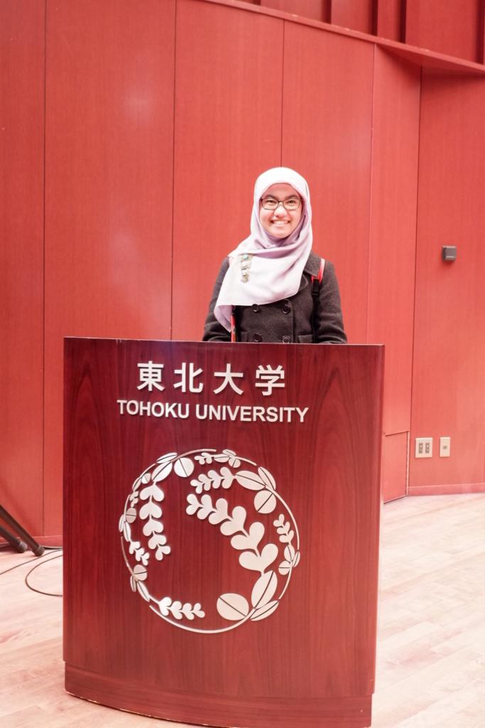
Sri Oktamuliani
@unand.ac.id
Physics Department / Faculty of mathematical and Natural science
Universitas Andalas
EDUCATION
Bachelor Degree - Universitas Andalas
Master Degree - Institut Teknologi Bandung
Doctor Degree - Tohoku University
Scopus Publications
Scholar Citations
Scholar h-index
Scholar i10-index
Scopus Publications
Sri Oktamuliani and Nurul Khaira Sabila
EDP Sciences
This study aimed to minimize radiation risk in Solok Selatan by analyzing active concentrations of 226Ra, 232Th, and 40K, calculating excess lifetime cancer risk (ELCR) from annual effective dose equivalent (AEDE). Soil samples from seven sites in the Solok Selatan, 0 – 5 cm deep, were tested with a high-purity germanium (HPGe) detector. 232Th concentrations exceeded the established global standard of 30 Bq/kg. In addition, the study included the determination of Radium Equivalent (Raeq), absorbed gamma dose rate (D), AEDE, and ELCR. Annual effective dose ranged from 68.33 to 19.92 μSv/y, below the global average of 80 μSv/y. The ELCR, the critical measure for understanding potential health risks, was 0.27 ± 0.12 x 10⁻³, under the global average of 0.29 x 10⁻³. Interestingly, areas closer to the geothermal sources, especially Koto Baru and Sungai Pagu, showed slightly higher natural radioactivity and corresponding effects. These findings emphasize the importance of rigorous surveillance and proactive measures in areas with high radioactivity characteristics. In summary, the results of this study provide important insights into the radiological landscape of Solok Selatan, urgently assisting in addressing potential health risks through informed risk assessment and strategies.
Sri Oktamuliani, Sri Rahayu Alfitri Usna, and Dinda Nurul Syifa
AIP Publishing
S. Oktamuliani, D. Fitriyani, R. Adrial, E. Sparzinanda, and Y. Saijo
AIP Publishing
The de-aliasing method is developed for Doppler velocity data of echocardiography. Physiological flow velocities exceeded low and high Nyquist velocity, resulting in aliasing. Aliasing occurs when the sampled signal is less than twice the highest frequency in the signal. The system does not take enough samples to ascertain which direction the flow occurs. Therefore, the scale and direction are displayed incorrectly in echocardiography. Echodynamography, which came as an idea of the limited information obtained from color Doppler echocardiography, is a method of estimating and visualizing three-dimensional (3D) blood flow velocity vectors two-dimensional (2D) observations plane by applying flow dynamics theory to the Doppler velocity in the heart. Color Doppler echocardiography is the premiere modality to analyze blood flow in clinical practice. The de-aliasing method is applied in Echodynamography. De-aliasing extends the Nyquist velocity range to complete ambiguous estimates of blood flow direction caused by aliasing. A sufficient velocity data de- aliasing scheme must be applied to recover the actual signal from the raw measurement. The method is tested with color Doppler echocardiography. The results show that the analytical study demonstrated that the de-aliasing method could efficiently and effectively reconstruct color Doppler velocity at the blood data point. Future discussion of the method limitations and possible improvements are needed.
Sri Oktamuliani, Naoya Kanno, Moe Maeda, Kaoru Hasegawa, and Yoshifumi Saijo
SAGE Publications
Echodynamography (EDG) is a computational method to estimate and visualize two-dimensional flow velocity vectors by applying dynamic flow theories to color Doppler echocardiography. The EDG method must be validated if applied to human cardiac flow function. However, a few studies of flow estimated have compared by EDG to the flow data were acquired by other methods. In this study, EDG was validated by comparing the analysis of estimating and visualizing flow velocity vectors obtained by original particle image velocimetry (PIV) based on a left ventricular (LV) phantom hydrogel (in vitro studies) and by EDG based on the virtual Doppler velocity. Velocity measured by PIV method and velocity estimated by EDG method in the perpendicular direction and the radial direction were compared. Regression analysis for the velocity estimated in the radial direction revealed an excellent correlation ([Formula: see text], slope = 0.96) and moderate correlation in the perpendicular direction ([Formula: see text], slope = 0.46). As revealed by the Bland–Altman plot, however, overestimations and higher relative error were observed in the perpendicular direction (0.51 ± 2.75 mm/s) and in the radial direction (–2.15 ± 21.13 mm/s). The percentage error of the norm-wise relative error of the velocity discrepancy is less than [Formula: see text], and velocity magnitude followed the same trends and are of comparable magnitude. These findings indicate that good estimates of velocity can be obtained by the EDG method. Therefore, the EDG method was appropriate for estimating and visualizing velocity vectors in clinical studies for higher measurement accuracy and reliability. The clinical in vivo application showed that the EDG method has the ability to visualize blood flow velocity vectors and differentiate the clinical information of vortex parameters both in normal and abnormal LV subjects. In conclusion, the EDG method has potentially greater clinical acceptance as a tool assessment of LV during the cardiac cycle.
Sri Oktamuliani, Kaoru Hasegawa, and Yoshifumi Saijo
IEEE
Echodynamography (EDG) is a computational method to deduce two-dimensional (2D) blood flow vector from conventional color Doppler ultrasound image by considering that the blood flow is divided into vortex and base flow components. Left ventricular (LV) vortices indicate cardiac flow status influenced by LV wall motion. Thus, quantitative assessment of LV vortices may become new and sensitive parameters for cardiac function. In the present study, quantitative parameters of LV vortices such as vortex index, vortex size, and Reynolds number were calculated and relation between each parameter was assessed. Six healthy volunteers and three patients with myocardial infarction (MI) who underwent color Doppler echocardiography (CDE) were involved in the study. Serial CDE images in apical three-chamber view were recorded and 2D blood flow vector was superimposed on the CDE image. Vortex index, vortex size, and Reynolds number were compared between the normal volunteers and the MI patients. The results showed that vortex index (3.09±2.06 vs. 3.34±2.33, p<0.05), vortex size (1.76 0.69 vs. 2.01 ±0.68, p<0.05), Reynolds number (1020±603 vs.±1312 1046, p<±0.05) were significantly greater in the MI patients than in the healthy volunteers. Vortex equivalent diameter in LV showed significant positive correlation with Reynolds number (R2 = 0.799, y = 0.001x + 0.7098, p < 0.05) in healthy volunteers and (R2 = 0.6404, y = 0.0005x+1.3185, p<0.05) in MI patients. Vortex index showed positive correlation with Reynolds number (R2 = 0.9351, y = 0.002x+0.1397, p<0.05) in healthy volunteers and (R2 = 0.758, y = 0.0019x+0.7957, p<0.05) in MI patients. In conclusion, EDG provides information on LV hemodynamics by quantitative LV vortices parameters both in healthy volunteers and MI patients.
Tadanori Minagawa, Yoshifumi Saijo, Sri Oktamuliani, Takafumi Kurokawa, Hiroyuki Nakajima, Kaoru Hasegawa, Takayuki Matsuoka, Takuya Shimizu, Makoto Miura, Takahiro Ohara,et al.
IEEE
Surgical intervention for aortic valve stenosis (AS) has been established; however its diagnosis based on echocardiographic assessment is still limited by aortic valvular velocity, aortic valvular pressure gradients, and color Doppler imaging. Echo-dynamography (EDG) is a method to determine intracardiac flow dynamics, such as two-dimensional blood flow velocity, vortex, and dynamic pressure. These flow dynamics may be influenced by left ventricular (LV) wall motion and the resistance in LV outflow caused by AS. The objective of the present study was to assess the changes and differences in LV vortices and vorticity before and after AS surgery. Five patients who underwent aortic valve replacement surgery for AS and five control patients were included. Besides routine echocardiographic measurement, EDG was applied to determine the two-dimensional blood flow vector and vorticity. The LV vortex flow in the isovolumetric contraction phase had multiple formations in preoperative cases. The clockwise vortex was found in all cases postoperatively; the vortex formation showed no significant difference between postoperative and normal control groups. EDG may serve important information on LV flow dynamics, non-invasively.
Sri Oktamuliani, Kaoru Hasegawa, and Yoshifumi Saijo
ACM Press
A study about the program of color Doppler to improve blood velocity estimation has been developed. Color Doppler echocardiographic images were acquired using Philips IE33 have a crucial issue the ambiguity of the offset beams when the correct velocity of the blood flow exceeds the velocity of the Nyquist limit. Therefore, velocity dealiasing is needed in quality control of color Doppler echocardiographic data to correct velocities using EDG (echodynamography) based on observer and machine interaction. The program is applied to echocardiography images of aortic stenosis subject as a technique to assess cardiovascular function. One challenging with color Doppler is correcting velocity aliasing. The observer determines the ambiguity area based on his or her knowledge. These results confirm the dealiasing and noise removing are vital elements in improving blood velocity estimation in cardiovascular diagnostic. Therefore, this study could use clinical tools based on color Doppler imaging.
Syahril Siregar, Sri Oktamuliani, and Yoshifumi Saijo
MDPI AG
We present a theoretical model of laser heating carbon nanotubes to determine the temperature profile during laser irradiation. Laser heating carbon nanotubes is an essential physics phenomenon in many aspects such as materials science, pharmacy, and medicine. In the present article, we explain the applications of carbon nanotubes for photoacoustic imaging contrast agents and photothermal therapy heating agents by evaluating the heat propagation in the carbon nanotube and its surrounding. Our model is constructed by applying the classical heat conduction equation. To simplify the problem, we assume the carbon nanotube is a solid cylinder with the length of the tube much larger than its diameter. The laser spot is also much larger than the dimension of carbon nanotubes. Consequently, we can neglect the length of tube dependence. Theoretically, we show that the temperature during laser heating is proportional to the diameter of carbon nanotube. Based on the solution of our model, we suggest using the larger diameter of carbon nanotubes to maximize the laser heating process. These results extend our understanding of the laser heating carbon nanotubes and provide the foundation for future technologically applying laser heating carbon nanotubes.
Sri Oktamuliani, Kaoru Hasegawa, and Yoshifumi Saijo
IEEE
The left ventricle has an important function in our body. The left ventricle receives oxygenated blood from the left atrium and brings through the aorta to the body. Therefore, estimation and visualization of blood flow velocity in the left ventricle are essential in clinical diagnosis. Color Doppler ultrasound is one of the instruments for left ventricle assessment that is used to detect blood flow velocity in the heart. However, color Doppler ultrasound shows the two-dimensional (2D) distribution of the one-dimensional (1D) velocity component of blood flow, forward or away along the transducer. The purpose of this study is to estimate and visualize the blood flow velocity in the left ventricle of the healthy volunteer and heart failure volunteers (mitral stenosis and mitral regurgitation) by Doppler ultrasound using echodynamography (EDG) method. The purpose of EDG is to estimate velocity component in a perpendicular direction to the ultrasound beam and to visualize the 2D velocity vectors of blood flow in the left ventricle. EDG composed the flow into the vortex (vortices laminar flow) and base flow (non-vortical laminar flows) components. The separation of flow is based on the continuity equation of fluid dynamics and hydrodynamics characteristics (stream function and flow function). The results show the blood flow velocity vectors are higher in the heart failure cases than the healthy case and indicated the vortex existence in the heart failure cases. In conclusion, the EDG method successfully estimated and visualized blood flow velocity in the left ventricle of the healthy and heart failure volunteers. Furthermore, the EDG method may contribute to the practical and clinical diagnosis of left ventricle assessment.
Sri Oktamuliani, Yoshifumi Saijo, and Kaoru Hasegawa
Acoustical Society of America
Blood flow dynamics were analyzed in healthy and myocardial infarction (MI) subjects using Echodynamography (EDG). EDG visualizes two-dimensional (2D) distribution of blood flow vector in the heart applying the theories of fluid dynamics to color Doppler echocardiography. The principle concept of EDG is that non-turbulent blood flow is described as the combination of base flow and vortex flow. The vortex flow is considered as a flow on a plane so that classical “stream function” is applied to obtain blood flow vector. The base flow is defined as the flow without vortex formation on the plane. Newly proposed “flow function” considers blood flow to and from other plane and the vector of the base flow is obtained with the flow function. Color Doppler data of heart apical three-chamber view were recorded by a commercially available medical ultrasound equipment. Blood flow distribution was different in healthy and MI subjects. In a healthy subject, vortex flow was observed as a whole blood flow in along the central flow area distributed to the base of the heart, and in the myocardial infarction the entire blood flow region the massive vortex flow was observed mainly at the apical part even in the ejection period.Blood flow dynamics were analyzed in healthy and myocardial infarction (MI) subjects using Echodynamography (EDG). EDG visualizes two-dimensional (2D) distribution of blood flow vector in the heart applying the theories of fluid dynamics to color Doppler echocardiography. The principle concept of EDG is that non-turbulent blood flow is described as the combination of base flow and vortex flow. The vortex flow is considered as a flow on a plane so that classical “stream function” is applied to obtain blood flow vector. The base flow is defined as the flow without vortex formation on the plane. Newly proposed “flow function” considers blood flow to and from other plane and the vector of the base flow is obtained with the flow function. Color Doppler data of heart apical three-chamber view were recorded by a commercially available medical ultrasound equipment. Blood flow distribution was different in healthy and MI subjects. In a healthy subject, vortex flow was observed as a whole blood flow in along the ce...
Sri Oktamuliani and Zaki Su’ud
AIP Publishing LLC
A preliminary study designs SPINNOR (Small Power Reactor, Indonesia, No On-Site Refueling) liquid metal Pb-Bi cooled fast reactors, fuel (U, Pu)N, 150 MWth have been performed. Neutronic calculation uses SRAC which is designed cylindrical core 2D (R-Z) 90 × 135 cm, on the core fuel composed of heterogeneous with percentage difference of PuN 10, 12, 13% and the result of calculation is effective neutron multiplication 1.0488. Power density distribution of the output SRAC is generated for thermal hydraulic calculation using Delphi based on Pascal language that have been developed. The research designed a reactor that is capable of natural circulation at inlet temperature 300 °C with variation of total mass flow rate. Total mass flow rate affect pressure drop and temperature outlet of the reactor core. The greater the total mass flow rate, the smaller the outlet temperature, but increase the pressure drop so that the chimney needed more higher to achieve natural circulation or condition of the system does no...

