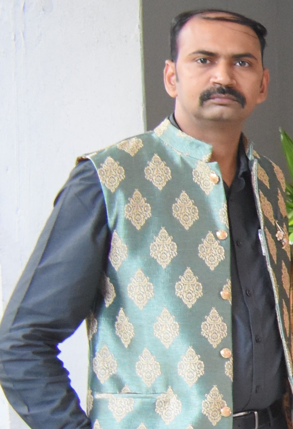
Mahendra Pratap Singh Tomar

@svvu.edu.in/college5

Assistant Professor-Veterinary Anatomy
Sri Venkateswara Veterinary University


https://researchid.co/anatomist

RESEARCH INTERESTS

Comparative Anatomy, Developmental Anatomy

13

Scopus Publications

148

Scholar Citations

8

Scholar h-index

7

Scholar i10-index

Scopus Publications

  • Anatomy of the reproductive system in the buck and doe
    Abhinov Verma, Mahendra P.S. Tomar, and Vikas Sachan

    Wiley

  • Enzyme histochemical characterization of orbital glands in fetuses of Indian buffalo (Bubalus bubalis)
    Mahendra Pratap Singh Tomar and Neelam Bansal

    PeerJ
    Background The orbital glands, viz. lacrimal gland, superficial and deep gland of third eyelid (LG, SGT and HG), are important for normal eye functions. These glands have different functions in various animals. The information about the enzyme histochemical nature of prenatal orbital glands in Indian buffalo seems to be unavailable. Therefore, the study was planned on orbital glands of six full term recently died fetuses from animals with dystocia. Methods The frozen sections of all these glands were subjected to standard localization protocols for Alkaline Phosphatase (AKPase), Glucose 6 phosphatase (G-6-Pase), Lactate dehydrogenase (LDH), Succinate dehydrogenase (SDH), Glucose 6 phosphate dehydrogenase (G-6-PD), Nicotinamide Adenine Dinucleotide Hydrogen Diaphorase (NADHD), Nicotinamide Adenine Dinucleotide Phosphate Hydrogen diaphorase (NADPHD), Dihydroxy phenylalanine oxidase (DOPA-O), Tyrosinase, non-specific esterase (NSE) and Carbonic anhydrase (CAse). Results The results revealed a mixed spectrum of reaction for the above enzymes in LG, SGT and HG which ranged from moderate (for LDH in SGT) to intense (for most of the enzymes in all three glands). However, DOPA-O, Tyrosinase and CAse did not show any reaction. From the present study, it can be postulated that the orbital glands of fetus have a high activity of metabolism as it has many developmental and functional activities which were mediated with the higher activity of the enzymes involved.

  • Endotoxin Biosynthesis: Genetics and Biochemistry of the Process


  • Endotoxins: A Basic Introduction


  • Anatomy of the vertebral column, ribs and sternum in orange rumped agouti (Dasyprocta leporina Linnaeus, 1758): Structural and Functional perspectives
    V. Sundaram, K. Jones, N. Mootoo and M. P. S. Tomar


    The axial skeleton of orange rumped agouti, Dasyprocta leporina, was studied for better understanding of its locomotor behaviour. The bones from eight adult agoutis of both sexes were observed for their anatomical features and functional significance. The vertebral formula was found to be C7T12L7S5Cy5–6. The well‐developed occipital crest, caudally oriented prominent axis spine and well‐developed transverse processes from C3–C7 indicated a highly flexible neck with greater sagittal mobility. Articular facets were horizontal in anterior series while oblique in the posterior series, which enabled them to perform both lateral and sagittal movements during locomotion. The caudally directed thoracic spines, T12 as anticlinal vertebra and prominent mamillary process in the posterior series were suggestive of strong dorso‐ventral flexion/extension and rotation. The robust lumbar vertebrae, well‐developed transverse processes with cranio‐ventral extension, were the feature for powerful sagittal/dorsoventral movement. The presence of spinous processes and well‐developed transverse processes in all caudal vertebrae was an indication of a highly movable tail. The ribs were 13 pairs with first seven as sternal and six as asternal. They were laterally compressed in the anterior series as a cursorial adaptation. A strong muscular attachment to vertebrae provides this rodent speed, agility, dexterity and strength suitable for survival in food chain.

  • Foetal embalming in buffalo-An umbilical approach


  • Prenatal development of retina in buffalo (Bubalus bubalis)
    Mahendra Pratap Singh Tomar and Neelam Bansal

    Wiley
    The development of retina in Indian buffalo (Bubalus bubalis) has not been reported previously. The aim of the present study was therefore to report the major landmarks and the time course in the development of retina. Serial histological sections of Indian buffalo embryos and foetuses were used as group1 (<20.0 cm CVRL), group2 (>20.0 but <40.0 cm CVRL) and group3 (>40.0 cm CVRL). Age estimation was made on the basis of crown vertebral‐rump length (CVRL), which ranged between 36 and 286 days (1.6–94.0 cm). The retina in Indian buffalo was developed in a similar manner to that of the other mammals with the principal differences in the time of occurrence of various layers of this nervous tunic. In 36 days (1.6 cm stage), the foetal retina was composed of pigmented layer and the layer of neuroblasts. Differentiation of layers was first observed in 47 days (4.0 cm CVRL) which became prominent in 52 days (5.1 cm stage). At 120 days (20.5 cm stage), the differentiation of inner plexiform layer and inner nuclear layer was evident. At 143 days (31.0 cm) foetal age, the faint line in neuroblastic layer was the first evidence of the future outer plexiform layer. In foetuses of group III, the retina was comprised of all 10 layers (eight cell layers and two membranes) viz. pigmented epithelium, layer of rods and cones, outer limiting membrane, outer nuclear layer, outer plexiform layer, inner nuclear layer, inner plexiform layer, ganglion cell layer, layer of nerve fibres and the inner limiting membrane.

  • Gross anatomy of scapula in tiger (Panthera tigris)
    M.P.S. Tomar, J.S. Taluja, Rakhi Vaish, A.B. Shrivastav, Apra Shahi, and Deepak Sumbria

    Agricultural Research Communication Center
    The present study was conducted on the scapula of five adult tigers to record the characteristic features of scapula bone. It was placed on lateral aspect of thorax, directed downward and forward. It was in the form of wide plate having scapular spine on lateral aspect. The height of spine increased gradually towards the distal end. The acromian process was subdivided into hamate process and suprahamate process. Hamate process overhanged the glenoid cavity. The suprahamate process was in the form of thin triangular plate directed backwards. The supraspinous fossa presented undulating surface in its middle. The infraspinous fossa was triangular and more or less flattened. Subscapular fossa was shallow and presented two prominent ridges. The caudal angle of the scapula was terminated in to glenoid cavity which was oval to quadrangular in shape. Cranial and proximal to the glenoid cavity prominent supraglenoid tubercle was observed which had hook shaped coracoid process. Scapula of both the sides were morphologically similar but the morphometrical values for the right scapula were non-significantly higher than the left counterpart (t less than 0.05), which may be of some biomechanical importance.

  • Enzyme histochemistry of extraocular muscles in buffalo fetuses


  • Morphometry of sternum of pigeon, crow and owl


  • Comparative gross morphometry of the hooves of chital (Axis Axis) and domestic goat (Capra Hircus)


  • Gross morphometrical studies of sternum of Pariah Kite (Milvus migrans)
    Tomar MPS, Vaish Rakhi, Parmar MK, and Shrivastav Yogita

    ScopeMed
    The sternum of an adult Pariah kite (Milvus migrans) was studied for its gross morphometry. It was procured from Department of Wildlife Health and Management. The sternum of Pariah Kite was in the form of quadrilateral plate with dorsal, concave surface and ventral, convex surface. It formed the thoracic floor and was directed backwards and downwards in an oblique manner. The length and width of sternum were 6.00 cm and 4.20 cm., respectively. Ventral projection, the carina was in the form of thin curved plate, the height of which decreased from before backwards. It was 6.00 cm long, 1.30cm wide and 0.30cm thick (at anterior end). Anterior border was triangular and had an elongated facet on either side for articulation with distal extremity of the coracoids. The caudolateral angles were prominent. At

  • Gross morphometrical studies on pectoral girdle of Pariah kite (Milvus migrans)
    M.P.S. Tomar, Rakhi Vaish, Nidhi Rajput, and A.B. Shrivastav

    Medwell Publications
RECENT SCHOLAR PUBLICATIONS

  • Anatomy of the Reproductive System in the Buck and Doe
    A Verma, MPS Tomar, V Sachan
    Elements of Reproduction and Reproductive Diseases of Goats, 1-12 2025

  • The Pectoral Limb of Pariah Kite (Milvus migrans): A Gross Morphometrical Study
    Y Pandey, A Pandey, AB Shrivastav, MPS Tomar, R Vaish
    BP International 2023

  • Enzyme histochemical characterization of orbital glands in fetuses of Indian buffalo (Bubalus bubalis)
    MPS Tomar, N Bansal
    PeerJ 11, e15196 2023

  • Cardiomegaly: A brief review with basic and physiotherapeutic approach
    BSTM P.S.
    Indian Journal of Physical rehabilitation 6 (1), 59-66 2022

  • COMPARATIVE ANATOMY OF WILD CARNIVORES
    MPSTDVVND Prasad
    Veterinary Anatomy On Multidisciplinary Mindscape 1, 58-65 2022

  • Humane innovations in veterinary anatomy education in India
    K Puppala, M Tomar, A Kantepudi, R Nuthalapaty, DP Dangeti, A Panniru
    Veterinaria 70 (Suppl 1), 33-44 2021

  • Anatomy of the vertebral column, ribs and sternum in orange rumped agouti (Dasyprocta leporina Linnaeus, 1758): Structural and Functional perspectives
    V Sundaram, K Jones, N Mootoo, MPS Tomar
    Anatomia, Histologia, Embryologia 50 (6), 985-995 2021

  • Comparative studies on different embalming fluids in ethically sourced canine cadavers.
    PVS Kishore, PSS Anusha, DD Prasad, K Archana, MPS Tomar, ...
    2021

  • Humerus of Indian Wildcat (Felis silvestris ornata: Gray, 1830)—A Gross Osteological Study
    D Palanisamy, MPS Tomar, PB Ankem, R Sekhar
    Int. J. Livest. Res 10, 49-54 2020

  • Foetal embalming in buffalo-an umbilical approach.
    PVS Kishore, MPS Tomar, K Archana, NKB Raju, D Prasad
    2019

  • Workshop Manual on Willed Body Programme and Humane Alternatives
    KAANDMPST P.V.S. KISHORE, N.K.B. RAJU
    NATIONAL WORKSHOP ON “WILLED BODY PROGRAMME AND HUMANE ALTERNATIVES FOR 2019

  • Perfusion and Insufflation Technique for Preservation of the Stomach and Intestine of a Large White Yorkshire Pig (Sus scrofa domesticus)
    PVS Kishore
    Indian Journal of Veterinary Anatomy 31 (1) 2019

  • Enzyme histochemistry of eyelids in buffalo fetuses (Bubalus bubalis).
    MPS Tomar, NB Neelam Bansal
    2019

  • Gross Anatomy of Femur in Royal Bengal Tiger (Panthera tigris)
    ABSDS Tomar, M.P.S., J.S. Taluja, Rakhi Vaish, Apra Shahi
    Indian Journal of Veterinary Anatomy 31 (1), 75-76 2019

  • Prenatal development of retina in buffalo (Bubalus bubalis)
    MPS Tomar, N Bansal
    Anatomia, Histologia, Embryologia 48 (2), 125-132 2019

  • Organ Embalming by Perfusion Technique: A Humane Alternative for Animal Sacrifice in Veterinary Education
    PVS Kishore, MPS Tomar, K Archana, NKB Raju, N Siva Rama Krishna
    Int J Livestock Res 9 (8), 237-42 2019

  • Macro-anatomy of Radius and Ulna in Indian Wildcat (Felis lybica ornata)
    RSU Dharani Palanisamy, M.P.S. Tomar, Prasanth Babu Ankem, S. Dhileswara Rao
    Indian Journal of Veterinary Anatomy 30 ((1)), 72-74 2018

  • GROSS ANATOMY OF RADIUS AND ULNA IN ROYAL BENGAL TIGER (Panthera tigris)
    ABSDS M.P.S. Tomar, J.S. Taluja, Rakhi Vaish
    Applied Biological Research 20 (2), 171-176 2018

  • Heart of Yellow Bellied Sea Snake (Hydrophis platurus): A Gross Morphological Study
    SAVPT Naveen Jonnalagadda, M.P.S. Tomar , Satish Putluru, Dharani Palanisamy
    International Journal of Current Microbiology and Applied Sciences 7 (3 2018

  • Light and Scanning Electron Microscopic Studies on Corneal Ontogeny in Buffalo
    NB M. P. S. Tomar
    ICAAP 2018 : International Conference on Animal Anatomy and Physiology 2018

MOST CITED SCHOLAR PUBLICATIONS

  • Morphometry of sternum of pigeon, crow and owl
    MA John, JS Sasan, K Ahmed, MPS Tomar, A Ahmad, AD Singh
    Indian Vet. J 91 (03), 40-41 2014
    Citations: 20

  • Comparative trichology of common wild herbivores of India
    HR Joshi, SA Gaikwad, MPS Tomar, K Shrivastava
    Advances in Applied Science Research 36, 3455-8 2012
    Citations: 16

  • Gross morphometrical studies of sternum of pariah kite (Milvus migrans)
    MPS Tomar, R Vaish, MK Parmar, AB Shrivastav, Y Tiwari
    Veterinary World 4 (4), 171 2011
    Citations: 15

  • Gross morphometrical studies on pectoral girdle of Pariah kite (Milvus migrans).
    MPS Tomar, RV Rakhi Vaish, NR Nidhi Rajput, AB Shrivastav
    2010
    Citations: 15

  • Gross anatomical study on humerus of tiger (Panthera tigris)
    MPS Tomar, JS Taluja, R Vaish, AB Shrivastav
    International journal of advanced research 2 (3), 1034-1040 2014
    Citations: 14

  • Gross anatomy of scapula in Tiger (Panthera tigris)
    MPS Tomar, JS Taluja, R Vaish, AB Shrivastav, A Shahi, D Sumbria
    Indian Journal of Animal Research 52 (4), 547-550 2018
    Citations: 11

  • Gross Morphometrical Studies on Pectoral Limb of Pariah Kite (Milvus migrans
    Y Tiwari, A Pandey, AB Shrivastav, MPS Tomar, R Vaish
    Annual Review & Research in Biology 1 (4), 111-116 2011
    Citations: 11

  • Avian esophagus: A comparative microscopic study in birds with different feeding habits
    MP Tomar, HR Joshi, PJ Ramayya, R Vaish, AB Shrivastav
    Int J Med Health Res 9 (8), 5-6 2015
    Citations: 8

  • Gross morphology of scapula in Indian wild cat (Felis silvestris ornate: Gray, 1830)
    D Palanisamy, MPS Tomar, PB Ankem, RS Ullakula, N Jonnalagadda, ...
    Int. J. Curr. Microbiol. Appl. Sci., 7 (4), 2473-7 2018
    Citations: 7

  • Extraction of genomic DNA from formalin fixed tissues of different wild avian species
    K Shrivastava, MS Thakur, MPS Tomar, AB Shrivastav, SNS Parmar
    Annals of Biological Research 3 (7), 3174-3177 2012
    Citations: 5

  • Humerus of Indian Wildcat (Felis silvestris ornata: Gray, 1830)—A Gross Osteological Study
    D Palanisamy, MPS Tomar, PB Ankem, R Sekhar
    Int. J. Livest. Res 10, 49-54 2020
    Citations: 4

  • Perfusion and Insufflation Technique for Preservation of the Stomach and Intestine of a Large White Yorkshire Pig (Sus scrofa domesticus)
    PVS Kishore
    Indian Journal of Veterinary Anatomy 31 (1) 2019
    Citations: 4

  • Comparative studies on different embalming fluids in ethically sourced canine cadavers.
    PVS Kishore, PSS Anusha, DD Prasad, K Archana, MPS Tomar, ...
    2021
    Citations: 2

  • Foetal embalming in buffalo-an umbilical approach.
    PVS Kishore, MPS Tomar, K Archana, NKB Raju, D Prasad
    2019
    Citations: 2

  • Prenatal development of retina in buffalo (Bubalus bubalis)
    MPS Tomar, N Bansal
    Anatomia, Histologia, Embryologia 48 (2), 125-132 2019
    Citations: 2

  • Organ Embalming by Perfusion Technique: A Humane Alternative for Animal Sacrifice in Veterinary Education
    PVS Kishore, MPS Tomar, K Archana, NKB Raju, N Siva Rama Krishna
    Int J Livestock Res 9 (8), 237-42 2019
    Citations: 2

  • Heart of Yellow Bellied Sea Snake (Hydrophis platurus): A Gross Morphological Study
    SAVPT Naveen Jonnalagadda, M.P.S. Tomar , Satish Putluru, Dharani Palanisamy
    International Journal of Current Microbiology and Applied Sciences 7 (3 2018
    Citations: 2

  • Enzyme histochemistry of extraocular muscles in buffalo fetuses
    MPS Tomar, N Bansal
    Indian Veterinary Journal 94 (11), 36-38 2017
    Citations: 2

  • Enzyme histochemistry of eyelids in buffalo fetuses (Bubalus bubalis).
    MPS Tomar, NB Neelam Bansal
    2019
    Citations: 1

  • Gross Anatomy of Femur in Royal Bengal Tiger (Panthera tigris)
    ABSDS Tomar, M.P.S., J.S. Taluja, Rakhi Vaish, Apra Shahi
    Indian Journal of Veterinary Anatomy 31 (1), 75-76 2019
    Citations: 1